| Schedule | Lectures | Seminars | Tests | Glossary | Cases | Index | Review | Search | Feedback | |
| Schedule | Lectures | Seminars | Tests | Glossary | Cases | Index | Review | Search | Feedback | |
Patient 1. Welcome to the gynecology clinic. Your first patient is a 21 year old GOPO with her LMP 6 weeks ago. She is noted to have ovarian enlargement (7cm) on pelvic exam. She has no pelvic pain or tenderness. You should:
|
Click on your choice: |
This patient most likely has a benign ovarian tumor. Common findings include:
 Click here for a larger view |
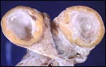
Follicular cyst.
Here is a benign cyst in an ovary. This is probably a follicular cyst.
Occasionally such cysts may reach several centimeters in size and, if they
rupture, can cause abdominal pain.
Credits: C. Matthew Peterson, M.D. |
 Click here for a larger view |
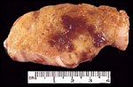
Corpus luteum cyst.
The corpus luteum secretes progesterone which
induces a secretory endometrium. It normally
regresses in 14 days unless it is rescued by
increasing concentrations of human chorionic
gonadotropin from a pregnancy.
Credits: Alan B.P. Ng |
 Click here for a larger view |
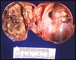
Endometrioma.
This is a section through an ovary to demonstrate several irregular
hemorrhagic areas of endometriosis. Sometimes the blood is darker and
gives the foci of endometriosis the gross appearance of "powder burns". Typical locations for endometriosis include: ovaries, uterine ligaments, rectovaginal septum, pelvic peritoneum, and laparotomy scars. Endometriosis may even be found at more distant locations such as appendix and vagina.
Credits: C. Matthew Peterson, M.D. |
 Click here for a larger view |
Mature cystic teratoma.
Teratomas, often called dermoid tumors, represent
25% of all benign neoplasms. The most common
elements are components of stratified squamous
epithelium, hence their name: dermoid tumor.
Credits: Alan B.P. Ng |
 Click here for a larger view |
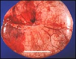
Serous cystadenoma.
These tumors comprise 20% of all ovarian neoplasms.
The epithelium is a single layer of regular
cuboidal epithelium, with basal nuclei and rare
mitoses.
Credits: Alan B.P. Ng |
 Click here for a larger view |
Mucinous cystadenoma.
These tumors comprise 20% of all neoplasms, and 50% of ovarian neoplasms found in women less than 20 years of age. They are frequently multiloculated. The favored hypothesis for their origin is metaplastic surface epithelium as they have a predominance of endocervical gland type epithelium.
Credits: Alan B.P. Ng |
An ultrasound study may further clarify the diagnostic possibilities.
![]()