1. The Cranial Nerves and the Circle Of Willis
Revised August 2, 2007
The objectives of this chapter are to identify:
- The structural divisions of the central nervous system (CNS).
- The cranial nerve roots associated with each brain stem division.
- The connective tissue wrappings, or the meninges, of the brain and spinal cord.
- The major arteries, veins and dural sinuses.
I. Regions of the CNS
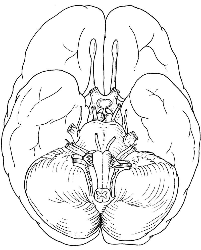
The neural tube finally consists of six divisions. Although each division has a characteristic appearance, both macroscopically and microscopically, it does not constitute a separate and independent functional unit. The central nervous system does not consist merely of a series of transverse slabs. From caudal to rostral, the divisions of the neural tube (#4281) are spinal cord, medulla (green), pons & cerebellum (yellow), midbrain (blue), diencephalon (purple), and cerebrum (uncolored). They are visible on the ventral surface of the brain as in fig 1a. Note: The labels on fig 1a and all figures in HyperBrain are linked to the glossary definition. To learn more as you go you can click on each label. However, the text explains many of the terms directly related to the chapter.
- spinal cord
- medulla oblongata = myelencephalon (#4985)
- pons and cerebellum = metencephalon (#4984)
- midbrain = mesencephalon (#4983)
- diencephalon, which consists mainly of the thalamus (#4269, light blue) and hypothalamus (dark blue)
- cerebrum = telencephalon, whose surface consists of the cerebral cortex (#8430).
Conveniently the divisions of the brain have the same names in the embryo and adult. Because of the tremendous development of the telencephalon dorsally and laterally, the midbrain and diencephalon cannot be seen externally except from the ventral surface.
II. The Cranial Nerves
A. Identify the cranial nerves:
Cranial nerves associated with the medulla (IX, X, XI, XII): Because the pons can be located easily, it serves as a good reference point (fig 1a). Caudal to the pons and tapering from it is the medulla, whose ventral surface is characterized by two pairs of elevations. The two medial elevations, one on each side of the midline furrow or ventral median fissure, are the pyramids (#5262). Lateral to each pyramid is the olive (#5261). Between the pyramid and olive of each side is a groove along which are attached the roots of cranial nerve XII (hypoglossal nerve) (#8443). This nerve innervates the striated muscles of the tongue. Does the right cranial nerve XII innervate the muscles on the left or right side of the tongue?
Cranial nerves IX (glossopharyngeal nerve) and X (vagus nerve) emerge lateral to the olive (#11686). They are not easily distinguished unless their ganglia are attached (#7281). Cranial nerve XI (spinal accessory nerve) originates from the spinal cord and lies lateral to the medulla (#4952). It is often torn off when the brain is removed from the skull.
Cranial nerves associated with the pons (V, VI, VII, VIII): Along the border of the pons and medulla, and in line with rootlets of XII, are the roots of cranial nerve VI (abducens nerve) (#5776). This nerve innervates the lateral rectus muscle of the eye. Does it innervate the ipsilateral or contralateral muscle?
In the angle between the lateral borders of the pons and medulla and against the backdrop of the cerebellum are several large nerve roots. They comprise cranial nerves VII (facial nerve) (#5577) and VIII (vestibulocochlear nerve) (#5578). The roots of VII are medial to those of VIII. Midway along the lateral border of the pons is the largest bundle of cranial nerve rootlets; they are cranial nerve V (trigeminal nerve), the sensory nerve of the face (#5579).
Cranial nerves associated with the midbrain (III, IV): Cranial nerve IV (trochlear nerve) (#11715) is twice unique in that
1. All of its fibers decussate within the midbrain. However, once the nerve emerges, it innervates the superior oblique muscle on the same side; i.e., the right trochlear nerve innervates the right superior oblique muscle.
2. Its fibers emerge from the dorsal surface of the brain (specifically, at the junction of the pons and midbrain). It can best be seen by gently pulling down on the cerebellum to expose the dorsal surface of the brain stem and then teasing away the overlying arachnoid and vessels.
Rostral to the pons are two large fiber bundles known as the cerebral peduncles (#5264). Together they form a "V" whose two arms meet at the rostral pontine border. They consist of fibers (axons) that originate in the cerebral cortex and pass through the brain stem to the spinal cord. They represent the ventral surface of the mesencephalon or midbrain. In the angle between them, and near the pons, emerges cranial nerve III (oculomotor nerve) (#8460). The temporal lobes and blood vessels can obscure them, but gentle prying should reveal them.
Cranial nerve II: Located between the cerebral peduncles is the ventral surface of the diencephalon. What is seen is the raised portion called the tuber cinereum (#11880), to which the pituitary gland is attached. This gland is not seen because it remains in the sella turcica when the brain is removed from the skull. However, the stalk of the gland can be seen (#4738). Two small, rounded elevations occur just caudal to the tuber cinereum; these are well named the mammillary bodies (#4737). The tuber cinereum is flanked rostrally and laterally by the optic chiasm and optic tracts, which are a caudal continuation of cranial nerve II (optic nerve)(#5345).
Cranial nerve I (olfactory nerve) is associated with the telencephalon. The rootlets that make up the olfactory nerve are torn from their insertions into the olfactory bulb when the brain is removed from the skull. The olfactory bulb (#4965) and olfactory tract (#4963) are readily recognized.
B. Functional components of the cranial nerves
The cranial nerves can be understood without resorting to rhymes or nonsense verse. With the exception of I and II, the cranial nerves are nothing more than modified spinal nerves. Each spinal nerve consists of
- a dorsal root (sensory) and
- a ventral root (motor) (#52090, labeled a, sensory, and b, motor)
The cell bodies of the dorsal root axons are located in a dorsal root ganglion, which is located outside the CNS. These cell bodies and their axons originate from neural crest cells. The cell bodies of ventral root axons, however, are located inside the CNS in the ventral horn gray matter (#6687). These cells are derivatives of neuroectoderm. Consequently, dorsal root axons grow into the CNS and ventral root axons grow out of it.
Cranial nerves (#4175) can be summarized by considering them in relation to the plan of a typical spinal nerve. Cranial nerves III, IV, VI, and XII are comparable to ventral roots. Cranial nerves V, VII, IX, and X consist of both dorsal and ventral root components and, thus, resemble a complete spinal nerve. Therefore, one would expect that these nerves would have the equivalent of dorsal root ganglia. They do. Where are these ganglia located? What is the name given to the "dorsal root" (or sensory) ganglion of VII (#7276)? Of V? (#7275) There are other ganglia in the head such as the otic ganglion, which is an autonomic ganglion. These, of course, are not homologues to dorsal root ganglia but are autonomic ganglion, similar to sympathetic chain ganglia, except that all cranial nerve autonomic ganglia are parasympathetic rather than sympathetic.
Cranial nerve VIII is homologous to a dorsal root and does not have a ventral root component. Cranial nerve II is developmentally a cerebral vesicle evagination, not a peripheral nerve. The microscopic anatomy of CN II is significantly different from that of CN VIII. What cells form the myelin sheaths around the axons in each of these nerves? What is the origin of the cells in each case? If the axons of X are interrupted along their peripheral course, they will probably regenerate. What is the possibility that axons in the optic nerve (II) will regenerate? Why?
III. The Meninges
The meninges (meninx = membrane) consist of three membranes. The outermost and thickest is the dura mater (literally "hard" or "tough mother"), also known as the pachymeninx (pachy = thick) (#51064). The dura is folded in the midline to form the falx cerebri (#11624), which incompletely separates the two cerebral hemispheres. Its other major fold is the tentorium cerebelli (#11625), which separates the cerebellum from the cerebral hemispheres. The tentorium ("tent") stretches over the top of the posterior cranial fossa (#5911). What sits on top of the tentorium? What is immediately below it? The free edge of the tentorium forms the tentorial incisure (#15239), also called the tentorial notch. The posterior and middle cranial fossae (#5910) are continuous with one another through this notch. Suggest the functional significance of the dural membranes. The dura mater of the brain is innervated by the trigeminal nerve (V) and spinal nerves C2-3. The dura is exquisitely sensitive to pressure and mechanical distortion. Suggest possible causes for such mechanical distortion. Stimulation of these sensory nerves results in certain forms of headache.
Covering the surface of the brain (and spinal cord, too) are two other meningeal membranes, the pia mater (#4923) and the arachnoid (#4924). Together they are called the leptomeninges (lepto = slender). The pia mater fits tightly over the brain, dipping into all the crevices and furrows (#5178). The arachnoid, however, is only loosely attached to the brain (#5616). The space between the pia and arachnoid is the subarachnoid space (#5927), in which lie the cerebral arteries and cortical (superficial cerebral) veins. This space also contains cerebrospinal fluid (CSF). Connecting the arachnoid to the pia are wispy connective tissue fibers that resemble spider webs; these give the arachnoid ("cobweb-like") its name. The subarachnoid space is enlarged in certain locations to form cisterns (#5617). These act like water-filled pillows that cushion the brain and allow it to "float over," as it were, the irregular bony contours of the cranial floor. The largest subarachnoid cistern, the cisterna magna (#12605), is located between the inferior surface of the cerebellum and the dorsal surface of the medulla. The cisterns communicate with one another, as well as with the rest of the subarachnoid space.
The cranial nerves pierce the meninges to leave the cranial cavity (#15245) and the spinal nerves pierce the meninges (#51286, #5400) to exit from the vertebral canal .
The meninges can become infected, as in meningitis (#5673) in which pus accumulates in the subarachnoid space. What effect could this have on the cranial nerves? The swelling due to the inflammation will stretch the dura, hence causing headache or pain, especially when the neck is flexed.
IV. The Vasculature
Two major arterial systems supply the brain, the internal carotid (#5905) system and the vertebrobasilar system (#5907). The internal carotid system supplies most of the cerebral hemispheres except for parts of the occipital and inferior temporal lobes. The vertebrobasilar system supplies the brain stem, including part of the diencephalon, and some of the cerebral hemispheres (i.e. the inferior surface of the temporal lobe and most of the occipital lobe). The distribution of the cerebral arteries is considered more fully in subsequent chapters.
The main arteries (fig 1b, fig. lc, NOTE: all labels on the figures are linked to the glossary terms) of the internal carotid system are the anterior (#4202) and middle cerebral arteries (#4201). The left and right vertebral arteries (#8447), located on each side of the medulla, unite at the junction of the medulla and pons to form a single midline artery called the basilar artery (#8450). This artery extends the rostrocaudal length of the pons. Branches of the vertebral and basilar arteries supply the medulla, pons, and cerebellum. Rostrally, at the junction between the pons and midbrain, the basilar artery bifurcates to form the left and right posterior cerebral arteries (#4198). The two major arterial systems are united by the posterior communicating arteries (#4199). The two anterior cerebral arteries are joined by the short anterior communicating artery (#4203), thus joining the left and right halves of the internal carotid circulation. This connection, plus the connection between the internal carotid and vertebrobasilar systems, forms the Circle of Willis. This circle of arterial anastomoses provides a margin of safety should one of the major arteries be obstructed.
Veins of the brain (fig 1d) drain into large collecting channels in the dural folds called dural sinuses. The veins that extend between the cortical (superficial cerebral) veins on the surface of the brain and the dural sinuses are bridging veins (#8433, #7979, #7964). The dural sinus along the superior edge of the falx cerebri is the superior sagittal sinus (#7971, #51064, #12420). Running along the inferior margin of the falx cerebri is the inferior sagittal sinus (#12541). It joins the great cerebral vein (of Galen) (#12547) to form the straight sinus (#12540), which runs in the midline along the border of the falx cerebri and tentorium cerebelli. This sinus meets the superior sagittal sinus at the internal occipital protuberance, where they form the confluence of the sinuses (#4103). Continuing laterally from the confluence on each side is a transverse sinus (#11620). It goes forward along the lateral edge of the tentorium to the sigmoid sinus (#52277, #7973), which drains into the internal jugular vein (#7301) after passing through the jugular foramen (#6953).
Click for the Syllabus Quiz for Chapter 1.
Movies
Self-Study
The following diagrams from Dr. David Morton can be used to examine yourself to see if you can name the arteries and cranial nerves.
| Syllabus Home | Chapter 2 |