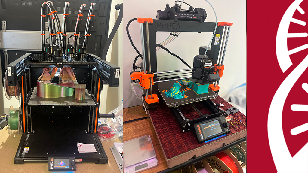
Located in the EHSEB 3100 computer lab, is a 3D printer lab, run by the Eccles Health Sciences Library. 3D printing services are available to University of Utah students, faculty, and staff across the health sciences campus. This program is currently free.
Meet the Maker of August: Hezro Da Silva is a third year medical student.
What did you make?
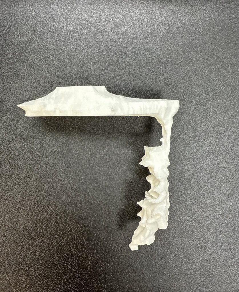
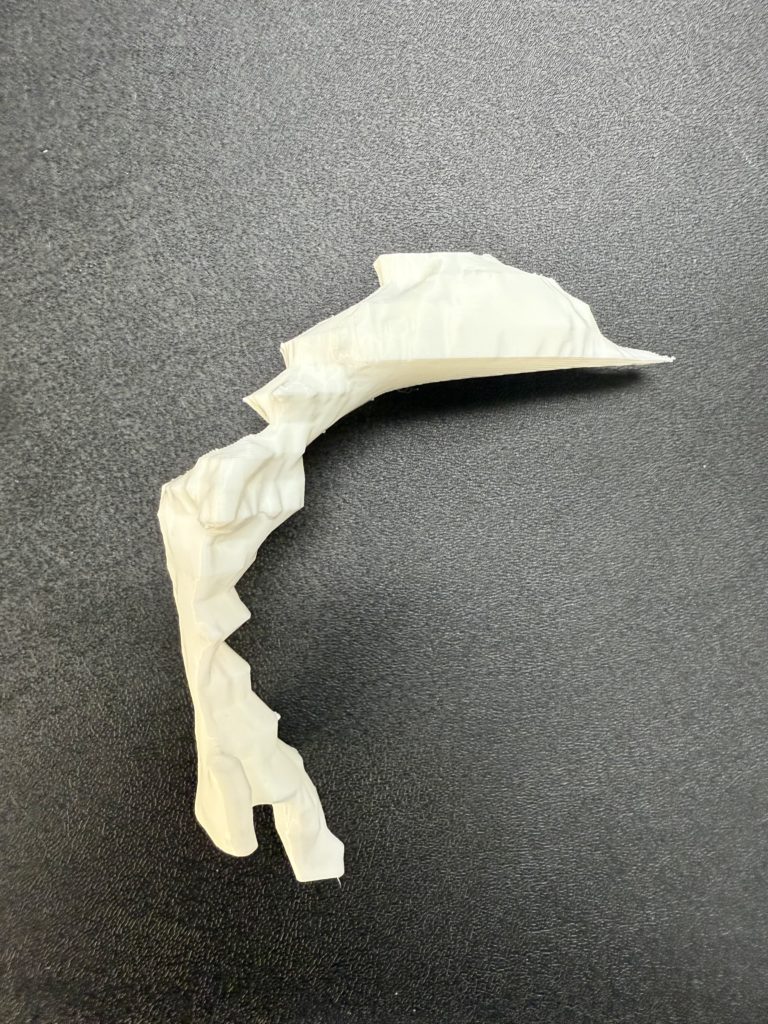
· We made two life-sized, 3D-printed models of the airspace within the human vocal tract—from the lips all the way down to the glottis (the space between the vocal folds).
Why did you make it?
· At the Voice Airway Swallowing Translational (VAST) Research Lab, one of our many ongoing studies explores two rare and debilitating neurogenic voice disorders: vocal tremor and laryngeal dystonia. These 3D models were an excellent way to visualize and compare anatomical differences in the vocal tract—specifically at the glottis—between disorders.
What was your process?
· We scanned participants with neurogenic voice disorders inside an MRI machine while they sustained the vowel “ah”. The scanner captured sequential slices, moving down from the lips to the glottis. Using Materialise Mimics, we converted the DICOM slices into masks of the vocal tract airspace at each level. Finally, these masks were compiled into full 3D models, which we were able to print in collaboration with our colleagues at the Eccles Health Sciences Library.
What was the hardest part of the process?
· Recruiting participants was the biggest challenge. These disorders are rare, and we often need to fly patients to Salt Lake City from across the country to participate in our studies.
What was your favorite part of the process?
· Finding that patients with vocal tremor have a significantly larger glottal cross-sectional area than those with laryngeal dystonia or healthy controls was an exciting part of the process. We even had an abstract on this topic accepted for a podium presentation at the American Society for Head and Neck Radiology conference in Fall 2025! Seeing the anatomical difference firsthand in these printed models was quite a rewarding moment.
How has this project contributed to your work or education?
· This project marks an exciting step toward possibly using MRI to improve diagnostic classification of complex neurogenic voice disorders.
What do you want to make next?
· One idea we are interested in is printing a life-sized, hollow model of the nose, mouth, and throat. This can serve as a hands-on training tool for Speech-Language Pathologists practicing nasoendoscopy—a procedure where a camera is inserted through the nose to visualize the vocal folds.
Start your own project!
Need help? ehsl-techies@lists.utah.edu