| Schedule | Lectures | Seminars | Tests | Glossary | Cases | Index | Review | Search | Feedback | |
| Schedule | Lectures | Seminars | Tests | Glossary | Cases | Index | Review | Search | Feedback | |
The following ovarian neoplasms are benign and are included in the differential diagnosis of ovarian enlargement. Dermoid cystic teratomas are the most common benign ovarian neoplasm.
 Click here for a larger view |
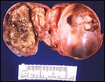
Mature cystic teratoma.
Teratomas, often called dermoid tumors, represent
25% of all benign neoplasms. The most common
elements are components of stratified squamous
epithelium, hence their name: dermoid tumor.
Credits: Alan B.P. Ng |
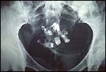
Dermoids make up 25% of all benign ovarian neoplasms. They are 80% of all benign ovarian neoplasms in young women found before the age of 20. The interior contains multiloculated cysts filled with bone, hair, sebum, and or teeth. They are occasionally detected on an x-ray of the abdomen because of the finding of a tooth in the abdomen.
 Click here for a larger view |
Radiograph of ovarian teratoma with teeth.
Teeth are occassionally part of the squamous
components of an ovarian teratoma.
Credits: Serono Laboratories |
Elements of all 3 germ layers (endoderm. mesoderm and ectoderm) can be found.
 Click here for a larger view |
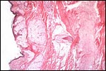
Mature cystic teratoma-microscopic.
Ectodermal tissue is usually most abundant and
represented by: squamous epithelium and
appendages, brain tissue, glia, retina, choroid
plexus, and ganglia. Mesodermal tissue is
represented by bone and cartilage.
Endodermal tissue is represented by
gastrointestinal and bronchial epithelium and
glands, thyroid, and salivary gland tissue.
Credits: Alan B.P. Ng |
The most common elements are stratified squamous epithelium and its appendages. Respiratory epithelium is found in 50-75% of the tumors. 10-25% are bilateral, and only 1-3% are malignant. The generally accepted theory for their origin is parthenogenic development. This origin is suggested because these tumors arise along the line of migration of the primordial germ cells at 4-6 weeks from the yolk sack to the primitive gonad. For this reason, these ovarian neoplasms are often classified as germ cell tumors.
Struma ovarii is a specialized form of a mature teratoma with a significant predominance of thyroid tissue or evidence of thyroid hormone activity from the tumor.
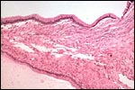
Serous cystadenomas comprise 20% of all ovarian neoplasms.
 Click here for a larger view |
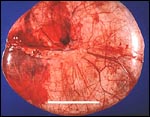
Serous cystadenoma.
These tumors comprise 20% of all ovarian neoplasms.
The epithelium is a single layer of regular
cuboidal epithelium, with basal nuclei and rare
mitoses.
Credits: Alan B.P. Ng |
Ten percent are bilateral. It is hypothesized that they arise through invaginations of the surface epithelium. They are large, unilocular and contain serous fluid. The epithelium is a single layer of regular cuboidal epithelium, with basal nuclei and rare mitoses.
Mucinous cystadenomas also comprise 20% of all ovarian neoplasms. In women less than 20 years of age, however, they constitute 50% of the benign epithelial neoplasms found. Five percent are bilateral. They are frequently multiloculated 15-30 cm cysts. The cyst has a single layer of tall columnar epithelial cells with mucin containing cytoplasm and basal nuclei.
 Click here for a larger view |
Serous cystadenomas.
A low cuboidal epithelium is present in the serous cystadenoma.
Credits: Edward C. Klatt, M.D. |
 Click here for a larger view |
Corpus luteum of the ovary, medium power.
The granulosa cells have undergone proliferation and alteration to lutein cells that produce progesterone. The lutein cells are large and polyhedral and the cytoplasm is foamy and eosinophilic.
Credits: Edward C. Klatt, M.D. |
Because a predominance of endocervical gland type epithelium is noted, a metaplastic surface epithelium is the favored hypothesis for their origin. Pseudomyxoma peritonei is seen in 2-5% of mucinous tumors. This condition is a massive collection of mucinous "slime" that accumulates in the peritoneal cavity. Some have suggested that this peritoneal lesion is an aggressive form of ovarian mucinous carcinoma of low malignant potential.
 View Video |
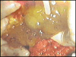
Pseudomyxoma peritonei. (VIDEO)
One of the complications of ovarian mucinous
cystadenomas is the development of pseudomyxoma
peritonei. This gelatinous accumulation may also be found associated with an appendiceal mucocele.
Occasionally all three entities are present making
the origin of the pseudomyxoma peritonei problematic. Histologically, the peritoneal implants of mucin secreting cells are a single
layer of mature cells filled with mucus and
basally arranged normal nuclei. This entity may
occur despite the absence of obvious rupture.
Care is taken during surgery on mucinous
cystadenomas to avoid spillage in the peritoneal
cavity.
Credits: C. Matthew Peterson, M.D. |
Other ovarian neoplasms include:
endometriod,
clear cell or mesonephroid,
Brenner
 Click here for a larger view |
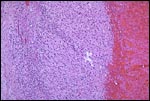
Brenner tumor.
Brenner tumors are comprised of solid to partly
cystic epithelial nests surrounded by stroma
composed of bundles of tightly packed spindle-
shaped cells. The epithelial cells are polygonal
and of squamoid type, with pale, eosinophilic
cytoplasm and oval nuclei with distinct nucleoli
and longitudinal grooving, which is commonly
described as "coffee-bean" in appearance.
Credits: Alan B.P. Ng |
stromal, and cystadenofibromas, and serous adenofibroma (solid variant of cystadenofibroma) tumors.
 Click here for a larger view |
Adenofibroma.
Dense,fibrous connective tissue with interspersed
glandular spaces
Credits: Edward C. Klatt, M.D. |
Evaluation of ovarian enlargement is best accomplished by ultrasound. Occasionally MRI or CT may be helpful if malignant neoplastic disorders are considered. CA 125 can be elevated in ovarian cancer, however benign situations like endometriosis and infection can also elevate this level.
![]()