| Schedule | Lectures | Seminars | Tests | Glossary | Cases | Index | Review | Search | Feedback | |
| Schedule | Lectures | Seminars | Tests | Glossary | Cases | Index | Review | Search | Feedback | |
Common non-neoplastic benign OVARIAN TUMORS include the following:
 Click here for a larger view |
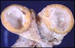
Follicle cysts.
Here is a benign cyst in an ovary. This is probably a follicular cyst.
Occasionally such cysts may reach several centimeters in size and, if they
rupture, can cause abdominal pain.
Credits: C. Matthew Peterson, M.D. |
 Click here for a larger view |
Corpus luteum cyst.
The corpus luteum secretes progesterone which
induces a secretory endometrium. It normally
regresses in 14 days unless it is rescued by
increasing concentrations of human chorionic
gonadotropin from a pregnancy.
Credits: Alan B.P. N |
 Click here for a larger view |
Mature cystic teratoma (dermoid).
Teratomas, often called dermoid tumors, represent
25% of all benign neoplasms. The most common
elements are components of stratified squamous
epithelium, hence their name: dermoid tumor.
Credits: Alan B.P. Ng |
 Click here for a larger view |
Serous cystadenoma.
These tumors comprise 20% of all ovarian neoplasms.
The epithelium is a single layer of regular
cuboidal epithelium, with basal nuclei and rare
mitoses.
Credits: Alan B.P. Ng |
 Click here for a larger view |
Mucinous cystadenoma.
These tumors comprise 20% of all neoplasms, and 50% of ovarian neoplasms found in women less than 20 years of age. They are frequently multiloculated. The favored hypothesis for their origin is metaplastic surface epithelium as they have a predominance of endocervical gland type epithelium.
Credits: Alan B.P. Ng |
 Click here for a larger view |
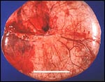
Endometrioma.
This is a section through an ovary to demonstrate several irregular
hemorrhagic areas of endometriosis. Sometimes the blood is darker and
gives the foci of endometriosis the gross appearance of "powder burns". Typical locations for endometriosis include: ovaries, uterine ligaments, rectovaginal septum, pelvic peritoneum, and laparotomy scars. Endometriosis may even be found at more distant locations such as appendix and vagina.
Credits: C. Matthew Peterson, M.D. |
 |
Pregnancy luteoma.
|
 Click here for a larger view |
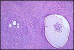
Theca-lutein cysts.
Theca lutein cysts are dominated by theca
interna cells. Grossly the cut surface of the
ovary is often partly yellow and partly
hemorrhagic. Hyperplasia of the theca interna
cells is the predominant characteristic. It is
believed that these cysts are the result of
excessive stimulation of the theca interna by
high levels of circulating hCG. Conditions
which high levels of hCG are found are multiple
pregnancies, gonadotropin therapy for infertility,
and trophoblastic disease.
Credits: Edward C. Klatt, M.D. |
The most common cause of bilateral ovarian enlargement is sclerocystic ovaries associated with PCOS
Other non-neoplastic solid benign tumors include:
 Click here for a larger view |
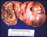
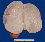
Fibroma.
This is the cut surface of a fibroma. Such neoplasms slowly enlarge
over the years.
Credits: C. Matthew Peterson, M.D. |
 Click here for a larger view |
Adenofibroma.
Dense,fibrous connective tissue with interspersed
glandular spaces
Credits: Edward C. Klatt, M.D. |
 Click here for a larger view |
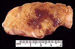
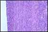
Ovary, Brenner tumor.
Solid and partially cystic epithelial nests are surrounded by a stroma composed of bundles of tightly-packed spindle shaped cells. The epithelial cells are polygonal and of the squamoid type, with pale, eosinophilic cytoplasm and oval nuclei having distinct nuclei and longitudinal grooving, a "coffee-ben appearance.
Credits: C. Matthew Peterson, M.D. |
![]()